|
 |
|
|
|
 |
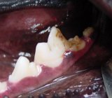
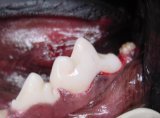
Enamel defect of lower left first molar. Abnormally shaped second molar. The third molar apparently is missing. |
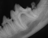
Pre-op xray to determine condition of first and second molars. Second molar is abnormal. Third molar is present but impacted. |
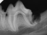
Post operative xray of extracted second and third molars. |
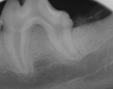
Post-op xray showing placement of synthetic bone material (Consil). |
Finished composite restoration of lower first molar. This case was submitted by Dr. Bert Dodd of Austin, Texas. |
[ Officers ] [ Find A Member ] [ Home ]
[ Contact Us ] [ Contact Webmaster ] [ Top of page ]
Copyright © 2002
Academy of Veterinary Dentistry
All rights reserved